Readers researching diabetic retinopathy screening need practical distinctions, reliable sources and a clear next step. The answer often depends on the circumstances, what can be verified and which professional or official instructions apply. This eye health guide focuses on the decision points people can actually investigate, rather than promising a universal result.
A practical health guide should help readers recognize what can be observed and what requires professional evaluation. It cannot diagnose an individual or promise a recovery date. The aim is to make symptoms, safety choices and next steps easier to discuss with a qualified clinician.
Why diabetic eye disease may have no early warning
Diabetic retinopathy screening matters because damage to retinal blood vessels can develop before a person notices a change in vision. The National Eye Institute advises people with diabetes to obtain comprehensive dilated eye examinations regularly, often at least annually. An eye doctor may recommend a different schedule based on the type of diabetes, eye findings, pregnancy or other circumstances. Symptoms alone are not a safe screening plan. Health advice depends on symptoms, personal history, examination findings, and professional assessment. A general guide cannot diagnose a condition or replace care. New or rapidly worsening symptoms, significant pain, or sudden functional changes deserve prompt medical attention rather than an extended self-treatment experiment. For further context, review Bridgemill Eye Care articles.
The retina is the light-sensitive tissue lining the back of the eye. Diabetes can affect its fine blood vessels, sometimes causing leakage, swelling or abnormal new vessel growth. Not everyone with diabetes develops the same disease pattern, and an ordinary eyeglass prescription check does not necessarily assess the retina fully. A dilated exam allows a clinician to look for changes that may otherwise be missed. One measurement or a single good day can be misleading. Clinicians often look for patterns across time and consider medications, previous injuries, workload, and other health conditions. Write down relevant changes in ordinary language so the visit focuses on the problem rather than a perfect self-diagnosis.
The retina and its small blood vessels
Early diabetic retinopathy may cause no obvious symptoms. Later problems can include blurred vision, spots, floaters or areas of missing vision, although these signs have other possible causes. Sudden vision loss, a new curtain-like shadow or abrupt major changes require urgent medical attention. Do not wait for the next routine screening when a warning sign appears. If a recommended exercise or routine increases symptoms substantially, stop and ask a qualified clinician how to modify it. Progress is seldom perfectly linear. A safe plan should specify both what to attempt and which symptoms mean the plan needs reassessment.
Vision changes are not a reliable screening method
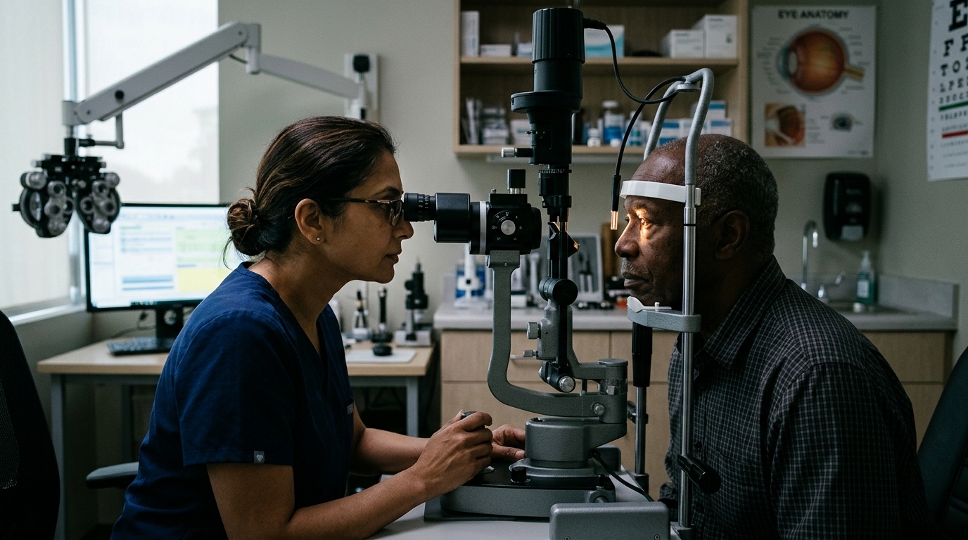
At a dilated eye exam, drops widen the pupil so the clinician can inspect more of the retina. The visit may also include visual acuity, eye pressure, imaging or other tests based on findings. Dilation can temporarily affect near vision and light sensitivity, so ask whether someone should drive you home. Follow the office’s specific preparation instructions. Commercial devices and viral techniques can promise certainty that research has not established. Read the intended use, limitations, and professional guidance before treating a product as a substitute for evaluation. A newer tool is not automatically better for every person.
Ask about OCT and retinal photographs
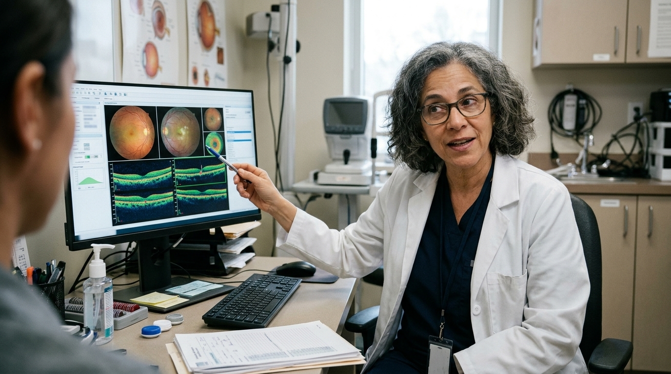
Optical coherence tomography, commonly called OCT, provides detailed cross-sectional images that can help detect retinal swelling. Retinal photographs document appearance and may assist comparison over time. Neither tool should be portrayed as a complete replacement for professional examination in every situation. Testing is selected according to symptoms and the clinician’s assessment. The practical goal is understanding, not fear. Identify what can be observed safely at home, which questions belong at an appointment, and what records will help with follow-up. Education is most useful when it makes appropriate care easier to access.
People who use insulin, tablets or other glucose-management tools should not change treatment based solely on an eye exam result. Eye findings provide one part of a larger health picture. Coordinate follow-up with the professionals managing diabetes and blood pressure, and ask for a written summary of significant ophthalmic findings.
Symptoms that need prompt attention
Blood sugar, blood pressure and cholesterol management contribute to long-term eye health, but good numbers do not eliminate the need for screening. Share your recent diabetes history, medications, prior eye conditions and any pregnancy-related concerns. A clinician may coordinate care with the diabetes team when findings suggest a need for closer systemic management. Risk factors describe probabilities across groups, not destinies for individuals. An athlete or patient may improve with a simple adjustment, require longer rehabilitation, or need another assessment. Avoid comparing your timeline with a social-media success story.
Patients may notice that their glasses prescription changes as glucose levels fluctuate. A temporary blur does not prove retinopathy, but it still deserves discussion. The retina needs assessment independently of how well a new pair of glasses seems to work.
What happens at a comprehensive dilated eye exam

If an examination identifies changes, the next step depends on severity. Options may include closer observation, treatment of diabetic macular edema, injections, laser procedures or other specialist care. Not everyone needs an immediate procedure. Ask what finding is being monitored, which symptoms should trigger a call, and when the next visit is recommended. A good professional discussion includes the expected benefits, burdens, cost, and alternatives of each option. Ask how success will be measured and what the plan is if symptoms persist. Shared decisions are more realistic than assuming one approach fits everyone.
What screening results can lead to
Keep the date of the last dilated exam and any imaging results with your general medical records. If your insurance plan or clinic changes, request a summary so new clinicians can compare findings. A shared timeline avoids guessing whether a new spot in a photograph represents progression or a difference in image quality. Keep instructions and questions together: current symptoms, activities that provoke them, prior tests, relevant medications, and the next planned review. Do not start, stop, or change a prescription based on an article. Clarify uncertainties with your treating professional.
Prepare medications and diabetes history
A person may read clearly on a vision chart and still have early retinal disease. Conversely, blurry vision can come from dry eye, cataract or a changing glasses prescription. That is why a comprehensive examination matters: it evaluates possible causes instead of attributing every symptom to diabetes or assuming comfortable vision means the retina is healthy. There is value in steady, proportionate action. Protect the basics of recovery, notice meaningful changes, and obtain individualized guidance when warning signs appear. The purpose of this guide is to help you ask better questions, not to make a diagnosis.
Follow the recommended screening interval
If cost or transportation makes appointments difficult, ask the care team about accessible clinics, insurance coverage and reasonable follow-up options. Retinal screening only protects vision when abnormal findings are reviewed and the recommended next steps occur.
Retinal health is best treated as part of routine diabetes care rather than an isolated one-time test. A documented screening schedule, review of findings and clear referral plan make it easier to act when change is detected. Ask the eye-care professional what was seen and when follow-up should occur, instead of leaving without understanding the next step.
Questions for your appointment: What findings support the working diagnosis? Which symptoms should prompt urgent contact? How will improvement be measured? Which activities or medicines need professional supervision? A written answer to each question can make follow-up care easier to understand and safer to carry out.
For authoritative context, consult National Eye Institute diabetic retinopathy. Its scope and limitations should be considered alongside the specific facts; the link is not a substitute for professional or local instructions.
The useful next step is a proportionate plan: document what you notice, follow credible professional instructions, and arrange reassessment if symptoms worsen or progress stalls. Education should improve questions and decisions, not substitute for an examination. Seek urgent help when serious warning signs occur.